Performance
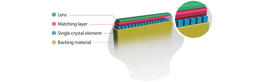
3T Transducer Technology with Single Crystal
Providing sharper images, all probes compatible with the M9 come equipped with Mindray’s unique 3T transducer technology. Enhanced with the addition of single crystal technology, M9 offers better penetration and color dynamic flow, especially during difficult-patient scanning.
Echo Boost™
Mindray’s unique adaptive signal processing technology with intelligent echo detection, designed to utilize the native signal-to-noise information to enhance the weak echo signals while suppressing the surrounding clutter noise, providing more balanced image brightness and improved visualization of myocardium tissue layers.
Tissue Tracking with Quantitative Analysis
The TT QA functionality on M9 allows for a simple, quick and non-invasive solution for the evaluation of left ventricular wall motion abnormalities.
LVO with Stress Echocardiography
M9’s premium capabilities allow for LV opacification during stress, enhancing discrimination between myocardial tissue and blood pool, providing better visualization of the endocardial surface.
PSHI™ (Phase Shift Harmonic Imaging)
Purified Harmonic Imaging for better contrast resolution providing clearer images with excellent resolution and less noise
Tissue Harmonic Imaging (THI)
Utilizing second harmonics generated from tissue boundary layers, THI significantly enhances contrast resolution and improves image quality especially for technically difficult subjects.
Tissue Specific Imaging (TSI)
Tissue Specific Imaging optimizes the image quality based on the properties of the tissue being scanned. Four imaging options are available including general, muscle, fluid and fat.
iBeam™
Permits use of multiple scanned angles to form a single image, resulting in enhanced contrast resolution and improved visualization
iClear™
Gain improved image quality based on auto structure detection
- Sharper & Continuous Edges
- Smooth Uniform Tissues
- Cleaner ‘no echo areas’
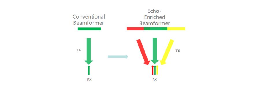
Echo-enriched Beam Forming
Echo-enriched beam former permits the use of traditionally neglected echo signals of adjacent beams to form one finer and stronger imaging beam, providing better ‘out-of-focus’ image resolution and deeper image penetration.
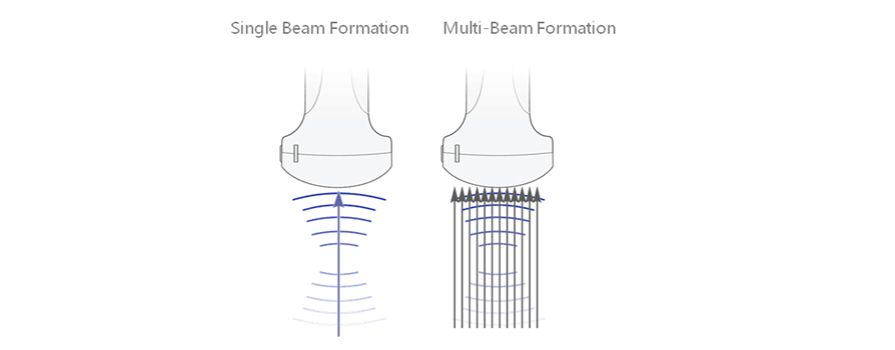
Multi-Beam Formation
Maximum 12 times tasking for one transmitted beam, resulting in excellent time resolution and higher frame rate.
Free Xros M™
Accurately evaluate myocardial motion at different phases, and simultaneously determine myocardial synchronization. High frame-rate providing you with accurate results
Auto EF
One intelligent way to analyze 2D echo clips to automatically recognize diastole/systole frames and output EDV/ESV/EF etc. results by Simpson method .
TDI (Tissue Doppler Imaging)
Tissue Doppler Imaging allows you to quantitatively evaluate local myocardial movement and function, providing complete TDI modes for faster and direct diagnoses.
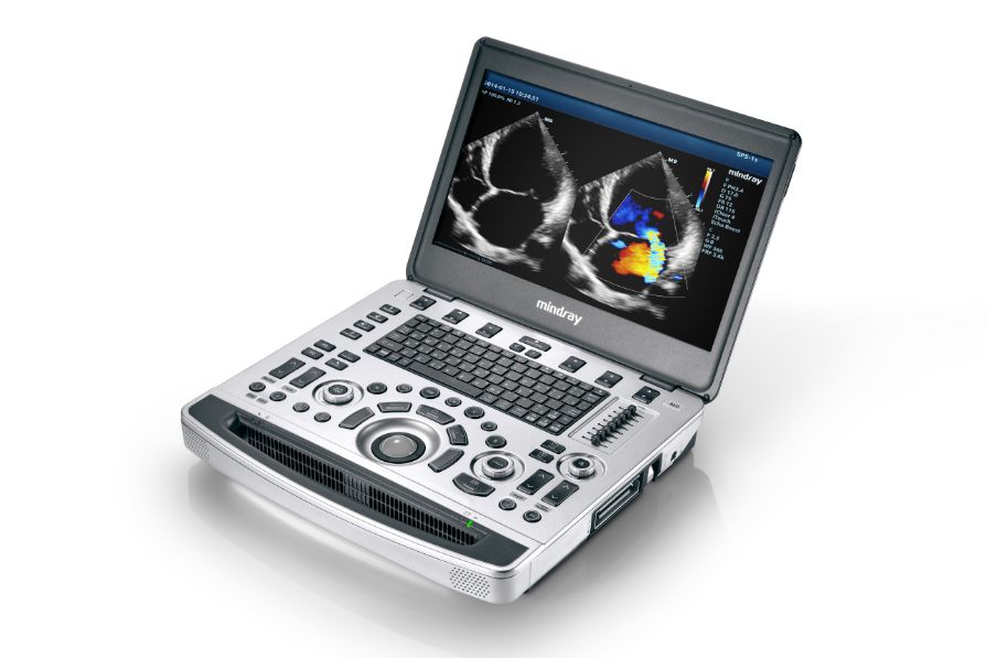
Ergonomics
Innovative crafted unit · Thin Magnesium-alloy body · 15.6” LED HD monitor with slim design · Built-in battery providing 90 min scanning time · High capacity SSD hard drive making patient data safer
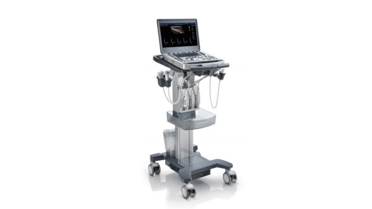
Customized Special Design Trolley · Inbuilt quick & easy locking system · iPower: over 3.5 hours scanning with trolley mounted battery pack
Green All the Way · Noiseless system · Automatic brightness adjustment · Reliable RoHS certified materials